Swine Flu Created In The University Of Massashutes Lab 242u19
This document was ed by and they confirmed that they have the permission to share it. If you are author or own the copyright of this book, please report to us by using this report form. Report l4457
Overview 6h3y3j
& View Swine Flu Created In The University Of Massashutes Lab as PDF for free.
More details h6z72
- Words: 7,474
- Pages: 8
JOURNAL OF VIROLOGY, Feb. 1992, p. 1066-1073
Vol. 66, No. 2
0022-538X/92/021066-08$02.00/0 Copyright © 1992, American Society for Microbiology
Hemagglutinin Mutations Related to Antigenic Variation in Hi Swine Influenza Viruses SHIUH-MING LUOH,"2t MARTHA W. McGREGOR,l AND VIRGINIA S. HINSHAW'12* Department of Pathobiological Sciences1 and Department of Medical Microbiology and Immunology,2 University of Wisconsin -Madison, Madison, Wisconsin 53706 Received 9 September 1991/Accepted 13 November 1991
Influenza A viruses are widespread in nature and cause disease in a variety of species, including humans, lower mammals, and birds (14). Influenza A viruses of the HlNl subtype were first detected in pigs in the United States in 1930. Such viruses continue to circulate in pigs and cause substantial disease problems, resulting in delayed marketing and increased expense for care and medication (7, 13). The mechanism for maintenance of the HlNi virus in pigs remains an unanswered question. It has been suggested that the viruses are maintained by age to young, susceptible pigs (13); therefore, the viruses may be subjected to little immune pressure and consequently undergo less antigenic variation than do human strains. To evaluate the level of antigenic variation of these viruses during their maintenance, we previously prepared a of monoclonal antibodies (MAbs) to the hemagglutinin (HA) of a recent HlNi swine virus (36). On the basis of the analysis of MAbselected escape mutants, we defined four antigenic sites, two of which overlap, on the Hi HA. When we used these MAbs to examine natural swine viruses isolated since 1965 from an enzootic area in Wisconsin, almost all of the viruses were highly conserved in these antigenic sites (36). Our next step, as described in this report, was to identify the genetic changes associated with the four antigenic sites on the HA of HlNi swine viruses by sequencing the HA genes of MAb-selected mutants and the parent strain, Sw/ IN/1726/88 (Sw/IN/88), for comparison with the Hi HA of AIPR/8/34 (6, 21, 32, 42). In our earlier studies, most natural swine viruses were shoWn to be antigenically conserved (36). In this study, we sequenced the HA genes of three representative swine
*
isolates from Wisconsin to examine their genetic relatedness. In addition, we previously observed that one of the natural swine isolates and a MAb-selected escape mutant had similar MAb reactivity (36), suggesting that their mutations were the same. To address this question, the sequence of the natural swine strain was obtained and compared with that of the MAb-selected mutants. Swine HlNl viruses also can be transmitted to other species, as evidenced by their ability to infect and cause disease in turkeys and humans (8, 15, 26, 34). Since 1980, turkeys have experienced disease outbreaks due to swine influenza viruses (15). Most importantly, humans are also susceptible to infection with swine viruses (8, 26, 34). Transmission of swine viruses to humans was first demonstrated in Wisconsin in 1976 (26); more recently, a woman in Wisconsin died from viral pneumonia after exposure to pigs with influenzalike disease at a fair (34). The causative agents in these human infections were serologically and genetically indistinguishable from contemporary swine viruses (26, 34). In addition, HlNi viruses, antigenically related to swine viruses, are present in ducks (3, 16). To understand the antigenic relationship of HlNi viruses, we previously used our MAbs to compare swine isolates with those from humans, ducks, and turkeys and found that two antigenic sites were conserved (36). In this study, HA sequence comparisons of swine, human, and duck isolates- also showed striking genetic similarities among HlNi viruses. In addition, we compared Hi and H5 HAs. Typically, there is no antigenic cross-reaction among distinct subtypes; however, serological and virus challenge studies suggest a low but biologically significant level of antigenic relatedness between the Hi and H5 HAs (1, 37). In examining this matter further, we compared the amino acid sequences of these HAs and their reactivities with anti-HA MAbs. Antigenic variation of swine Hi viruses obviously can and
Corresponding author.
t Present address, Amgen, Thousand Oaks, CA 91320.
1066
ed from jvi.asm.org by on April 26, 2009
The hemagglutinin (HA) of a recent swine influenza virus, A/Sw/IN/1726/88 (HlNl), was shown previously to have four antigenic sites, as determined from analysis of monoclonal antibody (MAb)-selected escape mutants. To define the HA mutations related to these antigenic sites, we cloned and sequenced the HA genes amplified by polymerase chain reaction of parent virus and MAb-selected escape mutants. The genetic data indicated the presence of four amino acid changes. After alignment with the three-dimensional structure of H3 HA, three changes were located on the distal tip of the HA, and the fourth was located within the loop on the HA. We then compared our antigenic sites, as defined by the changed amino acids, with the well-defined sites on the Hl HA of A/PR/8/34. The four amino acid residues corresponded with three antigenic sites on the HA of A/PR/8/34. This finding, in conjunction with our previous antigenic data, indicated that two of the four antigenic sites were overlapping. In addition, our previous studies indicated that one MAb-selected mutant and a recent, naturally occurring swine isolate reacted similarly with the MAb . However, their amino acid changes were different and also distant on the primary sequence but close topographically. This finding indicates that changes outside the antigenic site may also affect the site. A comparison of the HA amino acid sequences of early and recent swine isolates showed striking conservation of genetic sequences as well as of the antigenic sites. Thus, swine influenza viruses evolve more slowly than human viruses, possibly because they are not subjected to the same degree of immune selection.
VOL. 66, 1992
HEMAGGLUTININ MUTATIONS IN INFLUENZA VIRUS
does occur; however, in nature, the sites are quite conserved. We hope that by defining these conserved sites in our studies, we can better address the future control of these viruses.
with appropriate inserts were initially selected by probing with 32P-labeled cDNA obtained from Sw/WI/88 primed with Hi-specific primer as described previously (29). After identifying appropriate clones, we then cloned the HAl gene fragment into pSP65. We used a 32P-labeled transcript of the HAl gene cloned into pSP65 for detection of additional clones (24). The HAl gene fragment was cloned essentially as described above except that we used the reverse primer, H1-1065R (Fig. 1), to amplify the HAl gene fragments in PCR. Sequence of Hi HA genes. Plasmid DNAs from appropriate clones were extracted following a minilysis method (22). For sequencing, the double-stranded DNA was denatured to single-stranded DNA by treatment with 0.2 M NaOH and 0.02 mM EDTA (pH 8) at 37°C for 30 min. The alkalinedenatured DNA was then neutralized and precipitated by ethanol. DNA of both strands was sequenced by using a Sequenase kit (United States Biochemical Co.). We made a of synthetic oligonucleotides as sequencing primers which were either complementary to or of the same sense as viral RNA and which hybridized to the HAl region of the HA gene at intervals of 300 to 400 nucleotides (Fig. 1). To obtain the unambiguous and consensus sequence data and to eliminate the random mutations introduced by Taq polymerase during PCR, at least five clones from each HAl gene of different viruses were sequenced, and only the conserved mutations were reported. The sequence data were compiled, analyzed, and translated into deduced amino acid sequence by using the programs from the University of Wisconsin Genetics Center Group (UWGCG) (9). Mapping of mutations on Hl HA. To locate the mutations on the three-dimensional structure of Hi HA, we aligned sequence data of Hi HA with those of H3 HA. To do this, we first aligned the deduced amino acid sequence data of the Hi and H3 HAs, using the UWGCG program Bestfit (9). Three-dimensional alignment was obtained by using the method described previously (29). Pairwise identity comparisons of nucleotide and amino acid sequences of HA. Pairwise HAl nucleotide and amino acid sequence identity comparisons were performed with the UWGCG program Distances (9). Nucleotide sequence accession number. The nucleotide sequence presented in this article has been submitted to GenBank and given accession number M81707. RESULTS Sequence of the HA gene of Hi Sw/IN/88. To determine the sequence of the Hi HA gene of Sw/IN/88, we specifically amplified the HA viral RNA in one full-length segment by using a one-step PCR method followed by cloning into the pUC18 vector. To obtain the unambiguous sequence and compensate for any random mutations introduced by Taq polymerase during PCR (38), at least five clones were sequenced by the dideoxy-chain termination method. The plus-sense DNA sequence coding for the HA polypeptide is represented in Fig. 1, with the corresponding predicted amino acids listed above the nucleotide sequence. The Hi HA gene is 1,778 nucleotides in length and codes for a predicted protein with 17 amino acids in the signal peptide, 326 amino acids in the HAl polypeptide, and 222 amino acids in the HA2 polypeptide. In comparing the HA amino acid sequences of Sw/IN/88 and another Hi strain, A/NJ! 11/76 (4), the HAl and HA2 sequences are approximately 94 and 96.8% identical, respectively.
ed from jvi.asm.org by on April 26, 2009
MATERIALS AND METHODS Virus growth and purification. The viruses Sw/IN/88, five MAb-selected escape mutants of Sw/IN/88, Sw/WI/1915/88 (Sw/WI/88), SwIWII3/82 (Sw/WI/82), and SwlWI/46/76 (Sw/ WI/76) were from the influenza repository at the University of Wisconsin-Madison. For virus purification, viruses were propagated in 11-day-old embryonated hen eggs for 48 to 72 h at 35°C. The allantoic fluid was harvested and clarified by centrifugation at 7,000 rpm for 30 min. Polyethylene glycol (molecular weight, 8,000; Sigma Chemical Co., St. Louis, Mo.) was added to the allantoic fluid at a final concentration of 8% (wt/vol), stirred for 1.5 h on ice, and centrifuged at 7,000 rpm for 30 min. The resulting pellet was resuspended in STE buffer (0.1 M NaCl, 50 mM Tris hydrochloride [pH 7.8], 1 mM EDTA); viruses were purified by centrifugation on a 30 to 60% sucrose gradient at 24,000 rpm for 2 h in an SW28 rotor (Beckman Instruments, Inc., Fullerton, Calif.). The interface virus band was collected, diluted in STE, and repelleted at 24,000 rpm for 2.5 h in an SW28 rotor. The final virus pellet was resuspended overnight in STE buffer in the presence of 100 U of RNasin (Promega Biotec, Madison, Wis.) per ml. Viral RNA was isolated by treatment of purified viruses with proteinase K and sodium dodecyl sulfate as described previously (13), subjected to three phenol-chloroform (1:1) extractions and one chloroform extraction, and pelleted by ethanol precipitation. Cloning of HA genes. The primers (Fig. 1) used in cDNA synthesis, polymerase chain reaction (PCR), and dideoxychain termination sequencing were based on the published sequence of X-53A, a reassortant between A/PR/8/34 and A/NJ/ii/76 (4). The double-stranded DNA synthesis of the HA gene from Sw/IN/1726/88 was done by a one-step PCR method (30), which combines the first-strand cDNA synthesis and subsequent PCR amplification in a thermal cycler. Briefly, to anneal the primers to the template, approximately 1 Rxg of total viral RNA, 0.1 ,uM forward primer (Hl-7; Fig. 1), and 0.1 ,uM reverse primer (H1-1770R; Fig. 1), in a total volume of 20 RI, were heated at 65°C for 4 min and cooled to room temperature to allow annealing. The first-strand cDNA synthesis was done at 42°C for 45 min in a 100-pu reaction containing the preannealed 20-,u volume reaction, 6 pI of reaction buffer (1 M KCl, 0.6 M Tris hydrochloride [pH 8.3], 0.078 M MgCl2), 0.4 ,lI of 0.1 M DL-dithiothreitol, 6.4 RI of 10 ,uM deoxynucleoside triphosphates, 2.5 U of avian myeloblastosis virus reverse transcriptase (Pharmacia LKB, Piscataway, N.J.), and 2.5 U of Thermus aquaticus DNA polymerase (Taq polymerase; United States Biochemical Co., Cleveland, Ohio), overlaid with 100 pI of mineral oil. Sequentially, the same reaction was subjected to PCR amplification, which was done as follows: first cycle of 5 min at 93°C, 1.5 min at 52°C, and 3 min at 72°C; 29 cycles of 1.5 min at 930C, 1.5 min at 52°C, and 3 min at 72°C; and a final extension at 72°C for 10 min. The resulting PCR products were recovered from agarose gels by using GeneClean kit (Research Products International Corp., Mount Prospect, Ill.). The recovered products were kinase treated, blunt-end ligated into pUC18 vectors, transformed into Escherichia coli JM107 (11, 43), and selected on B medium (25) containing ampicillin, isopropyl-,-D-thiogalactopyranoside, and 5bromo - 4-chloro - 3 - indolyl- ,-D-galactopyranoside. Clones
1067
1068 1
51
J. VIROL.
LUOH ET AL. MetLysAlaIleLeuLeu AGCAAAAGCAGGGGAAAATAAAAGCCACCGAAATGAAGGCAATACTATTA _ Primer 7 HAl ValLeuLeuTyrThrPheThrAlaAlaAsnAlaAspThrLeuCysIleGl
GTCTTGCTATATACATTTACAGCCGCAAATGCAGACACATTATGTATCGG
951
LeuProPheGlnAsnIleHisProValThrIleGlyGluCysProLysTy CTCCCATTTCAGAATATACATCCAGTCACAATTGGAGAATGTCCAAAATA
1001
rValLysSerThrLysLeuArgMetAlaThrGlyLeuArgAsnIleProS TGTCAAAAGCACAAAATTGAGAATGGCTACAGGACTAAGGAATATCCCGT
E* A .
101
Primer 1009 W HA2 --
yTyrnisAlaAsnAsnSerThrAspThrValAspThrValLeuGluLysA TTATCATGCAAATAATTCAACTGACACTGTTGATACAGTACTAGAAAAGA Primer 121 mmm
151
snValThrValThrHisSerValAsnLeuLeuGluAspArgHisAsnGly ATGTAACAGTAACACACTCTGTTAACCTTCTAGAAGACAGACATAACGGA
201
1051
erIleGlnSer!rgGlyLeuPheGlyAlaIleAlaGlyPheIleGluGly
CTATTCAATCTAGAGGTCTGTTTGGAGCCATTGCTGGCTTTATTGAGGGG CCAGACAAACTTCGGTAACGA Primer 1065R
GlyTrpThrGlyMetIleAspGlyTrpTyrGlyTyrHiSHisGlnAsnGl 1101
GGATGGACAGGAATGATAGATGGATGGTACGGTTATCACCATCAAAATGA
AAACTATGTAAACTAAGGGGGGTAGCCCCATTGCATTTGGGTAAATGTAA
1151
uGlnGlySerGlyTyrAlaAlaAspArgLysSerThrGlnAsnAlaIleA GCAGGGATCAGGATATGCAGCTGACCGAAAGAGCACACAGAATGCCATTG
251
nIleAlaGlyTrpLeuLeuGlyAsnProGluCysGluLeuLeuPheThrA CATTGCAGGATGGCTCCTGGGAAACCCAGAATGTGAATTACTATTCACAG
1201
spGlyIleThrAsnLysValAsnSerValIleGluLysMetAsnThrGln ACGGAATCACTAACAAAGTAAACTCTGTTATTGAAAAGATGAACACACAA
301
CAAGCTCATGGTCTTACATTGTGGAAACATCTAACTCAGACAATGGGACA
LysLeuCysLysLeuArgGlyValAlaProLeuRisLeuGlyLysCysAs
laSerSerTrpSerTyrIleValGluThrSerAsnSerAspAsnGlyThr
PheThrAlaValGlyLysGluPheAsnHisLeuGluLysArgIleGluAs 1251
TTCACAGCAGTGGGTAAAGAATTCAACCACCTGGAAAAAAGAATAGAGAA
Primer
CysTyrProGlyAspPheIleAsnTyrGluGluLeuArgGluGlnLeuSe 351
TGTTACCCAGGAGATTTCATCAATTATGAAGAGCTAAGAGAGCAGTTGAG
nLeuAsnLysLysValAspAspGlyPheLeuAspValTrpThrTyrAsnA TTTAAACAAAAAGGTTGATGATGGTTTTCTGGATGTTTGGACTTACAATG Primer 1313
1351
laGluLeuLeuValLeuLeuGluAsnGluArgThrLeuAspTyrHisAsp CCGAACTGTTGGTTCTATTGGAAAATGAAAGAACTTTGGATTACCATGAC
1401
SerAsnValLysAsnLeuTyrGluLysValArgSerGlnLeuLysAsnAs TCAAATGTGAAGAACCTATATGAGAAAGTAAGAAGCCAGCTAAAAAACAA
1451
nAlaLysGluIleGlyAsnGlyCysPheGluPheTyrHisLysCysAspA TGCCAAGGAAATTGGAAATGGCTGCTTTGAATTTTACCACAAATGTGATG
1501
ACACGTGCATGGAGAGCGTCAAAAATGGGACTTATGATTACCCAAATTAC
345
rSerValSerSerPheGluArgPheGluIlePheProLysAlaSerSerT 401
CTCAGTGTCATCATTTGAAAGATTTGAGATATTCCCCAAGGCAAGTTCAT
451
rpProAsnHisGluThrAsnArgGlyValThrAlaAlaCysProTyrAla GGCCCAATCATGAAACGAATAGAGGTGTGACGGCAGCATGCCCTTATGCT
501
GGAGCAAACAGCTTCTACAGAAATTTAATATGGCTGGTAAAAAAAGGAAA
551
TTCATACCCAAAGCTCAGCAAATCCTATGTTAACAATAAGGAGAAGGAAG
Primer 474
GlyAlaAsnSerPheTyrArgAsnLeuIleTrpLeuValLysLysGlyAs
m
nSerTyrProLysLeLysSerTyrValAsnAsnLysGluLysGluV
spThrCysMetGlValLysAsnGlyThrTyrAspTyrProAsnTyr
alLeuValLeuTrpGlyIleHisHisProProThrSerThrAspGlnGln 601
651
701
751
SerLeuTyrGlnAsnAlaAspAlaTyrValPheValGlySerSerLysTy AGTCTCTACCAGAATGCAGATGCCTATGTTTTTGTGGGGTCATCAAAGTA Primer 685 rAsnLysLysPheLysProGluIleAlaThrArgProLysValArgGlyG
1551
TCAGAAGAATCAAAACTAAACAGAGAGGAAATAGATGGGGTAAAGCTGGA
1601
ATCAACAAGGATTTACCAGATTTTGGCGATCTATTCAACTGTCGCCAGTT
1651
erLeuValLeValSerLeuGlyAlaIleSerPheTrpMetCysSer CATTGGTACTGTCAGTCTCCCTGGGGGCAATCAGTTTCTGGATGTGCTCC
1701
AsnGlySerLeuGlnCysArgIleCysIle AATGGGTCTTTACAGTGCAGAATATGTATTTAAAACTAGGATTTCAGAGA
1751
Primer 1770R CATGAGAAAAAACACCCTTGTTTCTACT GTACTCTTTTTTGTGGGAAC
ThrArgIleTyrGlnIleLeuAlaIleTyrSerThrValAlaSerS
CAACAAGAAATTCAAGCCAGAAATAGCAACAAGACCCAAGGTGAGAGGTC lnAlaGlyArgMetAsnTyrTyrTrpThrLeuValGluProGlyAspThr AAGCAGGGAGAATGAACTATTACTGGACACTAGTAGAGCCTGGAGACACA
CTCT
IleThrPheGluAlaThrGlyAsnLeuValValProArgTyrAlaPheAl
851
ATAACATTCGAAGCAACTGGAAATCTAGTGGTACCAAGATATGCCTTCGC Primer 810 aMetLysArgGlySerGlySerGlyIleIleIleSerAspThrProValH AATGAAAAGAGGTTCTGGATCTGGTATTATCATTTCAGATACACCAGTCC
901
isAspCysAsnThrThrCysGlnThrProLysGlyAlaIleAsnThrSer ACGATTGTAATACGACTTGTCAAACACCCAAAGGTGCTATAAACACCAGC
801
SerGluGlLysLeuAsnArgGluGluIleAspGlyValLysLeuGl
TCCTCGTGCTATGGGGCATTCACCATCCACCTACCAGTACTGACCAACAA
mmm
mm m
FIG. 1. Nucleotide and deduced amino acid sequences of the HA gene of Sw/IN/1726/88, written in mRNA sense. Numbering starts at the first nucleotide at the 3' end of the gene. The N terminus of the mature HAl polypeptide and the cleavage point between the HAl and HA2 polypeptides are indicated with arrowheads. The solid rectangles represent potential glycosylation sites. The region on DNA to which primers (for cDNA synthesis, PCR, and sequencing) annealed are underlined and named. Reverse primers 1065R and 1770R, which hybridize to the complementary strand, are also represented.
Sequence analysis of HAl gene fragments from MAbselected escape mutants. In the previous work (36), we generated five MAb-selected escape mutants of Sw/IN/88, representing four antigenic sites, two of which overlap. Our goal in this study was to locate the genetic mutations in these MAb-selected escape mutants. Hereafter, we sequenced and analyzed only the HAl gene fragments, since the HAl polypeptide is the antigenically variable region of the HA molecule and the HA2 polypeptide is conserved (39-41). To determine the sequences, we amplified, cloned, and se-
quenced the HAl gene fragments by using the methods described above. The mutations of MAb-selected escape mutants were identified by comparing their sequences with that of the wild-type virus (Sw/IN/88). The positions and nature of the mutations are illustrated in Table 1. v2-15F1 had an alanineto-aspartic acid change at amino acid 156. The changes identified in v3F2c and v7Blb were the same, i.e., from lysine to glutamik acid; however, these occurred at different positions, i.e., amino acids 170 and 171, respectively. The
ed from jvi.asm.org by on April 26, 2009
1301
VOL. 66, 1992
HEMAGGLUTININ MUTATIONS IN INFLUENZA VIRUS
1069
TABLE 1. Mutations identified in MAb-selected variants of SW/ IN/1726/88 and their relationship to Hi human virus-A/PR/8/34
Variant
Nucleotide
Nucleotide change
Amino
Amino acid
v2-15F1
499 540 543 547 547
C to A A to G A to G G to A G to A
156 170 171 172 172
Ala to Asp Lys to Glu Lys to Glu Gly to Glu Gly to Glu
v3F2c
v7Blb v1-6B2 v4A12a
acid
change
Site related to human HA
Hi
Ca Sb Sa Sa Sa
FIG. 2. Locations of amino acid changes, identified in the MAbselected mutants of Sw/IN/1726/88 (U) and a naturally occurring swine virus (Sw/WI/1915/88) (0), relative to the H3 HA. The changed amino acids are marked, and the numbers shown are based on the Hi numbering. After alignment with the H3 HA sequence, amino acids 170, 171, 172, 156, and 86 correspond to amino acids 156, 157, 158, 142, and 78, respectively, on the H3 HA.
tides 84 to 1064) ranged from 94.4 to 99.9%, and the amino acid identities of mature HAl polypeptide (excluding signal peptide) ranged from 94.8 to 99.7%, indicating the high degree of relatedness among them. In addition, the predicted amino acid sequences of the entire HAl polypeptides were compared (Fig. 3). Sw/IN/88 was chosen as a standard with which other viruses were compared. Sw/WI/88 differed from Sw/IN/88 by 1 amino acid, from Sw/WI/82 by 11, and from Sw/WI/76 by 16, presumably as a result of accumulating amino acid changes over time. To determine whether the changed amino acids were restricted to antigenic sites analogous to the sites on the other Hi HA, we marked the equivalent sites on the amino acid sequence by alignment with AIPR/8/34 HA (6, 21, 32, 42) (Fig. 3). There were 16 widely scattered, changed amino acids on the HAs of swine viruses isolated from Wisconsin between 1976 and 1988. Three of sixteen changes were in antigenic sites, i.e., Val-to-Ala change at amino acid 90 in TABLE 2. Comparisona of MAb-selected variant of Sw/IN/1726/ 88 (v2-15F1) and Sw/WI/1915/88
v2-1SF1
Sw/WI/88 a
Comparison
499 289 was made in
Nucleotide change
Amino
C to A T to C
156 86
relationship
to
acid
Sw/IN/1726/88.
Amino acid
change Ala to Asp Leu to Ser
ed from jvi.asm.org by on April 26, 2009
same amino acid change, a glycine-to-glutamic acid shift at amino acid 172, was found in both v1-6B2 and v4A12a; this finding correlated with our previous results on MAb reactivity, showing that these two variants had changes in the same antigenic site. The previous antibody reactivity pattern suggested that v7Blb and two MAb-selected mutants (vl6B2 and v4A12a) may involve overlapping antigenic sites (36). The changed amino acid identified in v7Blb was at residue 171, which was adjacent to the changed amino acid 172 in v1-6B2 and v4A12a, further suggesting that amino acids 171 and 172 are in the same site. To relate these antigenic sites to the already defined sites on another Hi HA, we compared our sequences with that of A/PR/8/34 (6, 21, 32, 42). The parallel comparison revealed that amino acid 156 corresponds with antigenic site Ca, amino acid 170 corresponds with site Sb, and amino acids 171 and 172 correspond with site Sa (Table 1). Thus, these sequence changes in the swine HA correlate with three antigenic sites previously described for A/PR/8/34. These changed amino acids then were located on the three-dimensional structure of the HA molecule by alignment with H3 subtype of A/England/321M7 (12) and are topographically represented in Fig. 2. Amino acid 156 is in the loop of the HA, whereas amino acids 170, 171, and 172 are in the distal tip of the HA, bordering the proposed receptor binding site. Comparison of a recent, naturally occurring swine isolate and the MAb-selected mutant. We previously observed two reactivity patterns when naturally occurring swine isolates were tested in enzyme-linked immunosorbent assay (ELISA) and hemagglutination inhibition assay against our MAb (36). Most swine isolates reacted with all MAbs, while one isolate reacted with all but one MAb. The latter reactivity pattern was identified for a natural isolate Sw/WI/ 88, which had a MAb reactivity pattern similar to that of the MAb-selected mutant, v2-15F1. In determining whether the HA mutations of Sw/WI1/88 and v2-15F1 were the same, we sequenced the HAl gene fragment of Sw/WI/88 and compared the sequence with that of the variant (Table 2). The change in the HAl polypeptide of SwlWI/88 was from leucine to serine at amino acid 86, which was clearly different from the change in the mutant (v2-15F1), with an Ala-to-Asp change at amino acid 156. No other amino acid differences in HAl domains between the two were found, suggesting that these two changes are responsible for their antigenic phenotypes. Determination of genetic relatedness of Hi HAs from natural swine isolates. To define the level of genetic relatedness of Hi HAs of swine viruses isolated from Wisconsin pigs from 1976 to 1988, we sequenced the HAl gene fragments from a recent natural swine isolate (Sw/WI/88) and two earlier swine isolates (Sw/WI/82 and SwlWI/76). The pairwise nucleotide and amino acid identity comparisons of Sw/WI/88, Sw/WI/82, Sw/WI/76, and Sw/IN/88 are illustrated in Table 3. The nucleotide identities of HAl gene fragments (nucleo-
1070
J. VIROL.
LUOH ET AL. TABLE 3. Comparison of nucleotide and amino acid identities of HlNl influenza viruses No. of differences (% identity)a
Strain
Dk/Alb/35n6
NJ/116
Dk/Alb/35n76
Sw/WI/46/76
Sw/WI/3/82
Sw/WI/1915/88
Sw/IN/1726/88
231 (76.5) 30 (96.9)
226 (77.0) 47 (95.2) 40 (95.8)
227 (76.9)
226 (77.0) 58 (94.1) 54 (94.5) 46 (95.3) 1 (99.9)
230 (76.6)
NJ111M6
57 59 52 59 58
Sw/WI/46I76 Sw/WI/3/82 Sw/WI/1915/88 Sw/IN/1726/88
(82.5) (81.9) (84.1) (81.9) (82.2)
13 (96.0) 10 (96.6) 18 (94.2) 17 (94.5)
9 (97.2) 17 (94.8) 16 (95.1)
12 (96.3) 11 (96.6)
59 (94.0)
55 (94.4) 47 (95.2) 1 (99.7)
a Numbers of nucleotide differences (above the dashes) and amino acid differences (below the dashes) between two HAl gene fragments and two HAl polypeptides are listed. Values in parentheses denote the identity expressed as percentage of the gene. In this analysis, nucleotides 84 to 1064, which encodes mature HAl protein, are included and compared. Predicted amino acid residues of HAl proteins, excluding signal peptides, are compared. Sequence identity was determined by using the program Distances from UWGCG.
site Cb, Leu-to-Val change at 183, and Gly-to-Glu change at 187 in site Ca. These changes on the Hi HAs of the swine viruses, however, did not alter their reactivity with our MAb . One possible explanation is that conservative changes 1
HA1-
60
MKAILLVLLYTFTAANADTLCIGYHANNSTDTVDTVLEKNVTVTHSVNLLEDR}NGKLCK
WI/82 WI/76 NJ/76 Dk/ALB
C C E K F
AT AT
FC
VLK
I
V
61
IN /88 WI/88 WI /82 WI/76 NJ/76 Dk/ALB
osee
G I G IT G I
N
N I
Q
V
R
L V L N
D
V
K I
E
on
QLSSVSSFERFEIFPKASSWPNHETNRGVTAAYAGANSFYRNLIWLVgEGNSYPKLSK T
E
T
Dk/ALB
TK
K
181.
A
S S
S
L IT
T
AAA
SYVNINKEKEVLVLWGIHHPPTSTDQQSLYQNADAYVFVGSSKYNKKFKPEIATRPKVRGQ G
R R R RR A
G
L
NJ/76 Dk/ALB
G
G
T
V
241
IN/88 WI/88 WI/82
I
V CV
WI/76 NJ/76
IN/88 WI/88 WI/82 WI/76
12
S
121
IN/88 WI/88 WI/82
se
LRGVAPLHLGKCNIAGWLLGNPECELLFTASSWSYIVETSNSDNGTCYPGDFINYEELRE
A-
AGRMN
WI/76 NJ/7 6 Dk/ALB
SVSE
A A A A
S
N N N
I
IA W
LDQ
LNK
P
D
A A W A T A
EA2- W GAINTSLPFQNIHPVTIGEKYVKSTKLRMATGLRNIPSIQSRGLFGAIA
301
IN/88 WI/88 WI/82 WI/76 NJ/76 Dk/ALB
V
L S
V
I
V
FIG. 3. Amino acid comparison of the HAl domains of A/Sw! IN/1726/88, A/Sw/WI/1915/88, A/Sw/WI/3/82, A/Sw/WI/46/76, A/ NJ/11/76, and A/Dk/ALB/35/76. Only the amino acids different from those in the AISw/IN/1726/88 sequence are indicated. Numbering starts at the N terminus of HAl. Amino acid residues mapped in previously defined antigenic sites are shown: site Sa (D), site Sb (-), site Ca (A), and site Cb (0).
ed from jvi.asm.org by on April 26, 2009
IN/88 WI/88
involving noncharged amino acids, such as Val to Ala and Leu to Val, may have little impact on the antigenic sites. However, a charge change, such as Gly to Glu, might be expected to have an effect on the antigenic site, but none was detected by our MAbs. Genetic relatedness of HAs of Hi viruses from pigs, humans, and ducks. We determined the genetic relatedness of Hi HAs from swine viruses, viruses of swine origin transmitted to other species, and other Hi viruses. Because of the limited information on the sequences of Hi HAs, we have compared the published HA sequences of Dk/Alb/35/76 (Dk/Alb) (2) and of the virus reassortant A/NJ/11/76 (X-53A) with the sequences of four natural swine isolates that we determined. There was approximately 77% identity in HAl nucleotide sequence and 82% identity in amino acid sequence between Dk/Alb and other swine and human viruses. The HAl sequence identity of A/NJ/11/76 (X-53A) with the swine viruses was between 94 and 97%, and the amino acid identity was between 94 and 97% (Table 3). Apparently, the avian virus, Dk/Alb, is more distantly related to swine viruses, whereas AINJ/11/76 (X-53A) is more closely related. In addition, A/NJ/11/76 (X-53A) is more similar to the swine strain circulating in 1976 (Sw/WI/46/76) than to current swine viruses. The HAl amino acid comparison of A/NJ/11/76 (X-53A), Dk/Alb, and the natural swine isolates is shown in Table 3. We have previously shown that Dk/Alb did not react with MAbs M2-15F1 and M3F2c, which defined the antigenic sites Ca and Sb, respectively. When examining the location of changed amino acids with respect to Sw/IN/88, we found that 9 of 58 changes in Dk/Alb were located in the antigenic sites (Fig. 3); five (amino acids 154, 156, 159, 183, and 187) were located in antigenic site Ca, two (amino acids 173 and 203) were located in site Sb, and two (amino acids 88 and 91) were located in site Cb. Thus, the genetic mutations in antigenic sites Ca and Sb correlate with the lack of MAb reactivity to these sites. Similarly, the changes in antigenic site Cb were conservative and apparently not detected by our MAbs. In A/NJ/11/76 (X-53A), 4 of 17 amino acid changes were in the antigenic sites; they were amino acid 88 and 90 (in site Cb), 172 (in site Sa), and 187 (in site Ca). Because we had previously performed our antigenic studies on AINJ/8/76 (36) rather than A/NJ/11/76 (X-53A), we do not know whether these genetic mutations correlate with antigenic changes in A/NJ/11/76 (X-53A). Determination of relatedness of HAs between Hi and H5 viruses. Earlier studies had suggested that Hi and H5 HAs were related antigenically (1, 37); therefore, we compared the HAl amino acid sequences of Hi Sw/IN/88 and H5
VOL. 66, 1992
HEMAGGLUTININ MUTATIONS IN INFLUENZA VIRUS
DISCUSSION In this study, we located the antigenic sites on the Hi HA by sequencing the HAl gene fragments of Sw/IN/88 and its MAb-selected escape mutants. These antigenic sites, as defined by the single amino acid changes in MAb-selected mutants, were located within the loop and distal tip of the HA. To relate our antigenic sites to those already defined on the human H3 and Hi HAs, we aligned the sequences with those of A/Aichi/2/68 (X-31) (H3) (40) and A/PR18/34 (Hi) (6, 21, 33, 42). Amino acid 156 was in antigenic site Ca on Hi, which is equivalent to site A on H3. The three amino acid changes at 170, 171, and 172 correspond with sites Sa and Sb on Hi and with site B on H3. Although these genetic changes are located adjacent to each other, these variants can be distinguished with our MAb . Therefore, the combined antigenic and genetic data suggest that, as with A/PR/8/34, there are three antigenic sites on the Hi HA of Sw/IN/88. With the MAb , we previously reported that one MAb-selected mutant (v2-i5Fi) and a recent, naturally occurring swine isolate (Sw/WI/88) were antigenically similar in both hemagglutination inhibition assay and ELISA (36). However, the viruses had different mutations which were distant from each other on the primary amino acid sequence, i.e., 86 versus 156. When the changes were envisioned on the three-dimensional structure of HA, they were much closer to each other than the primary sequence would have indicated. Amino acid 86 was under, and in close proximity to, the antigenic loop which encomed amino acid 156 (antigenic site Ca). This finding suggests that the mutation at amino acid 86, outside the antigenic site, affects antibody recognition of the antigenic loop. Similar findings on foot-and-mouth disease virus have been reported by Parry et al. (28). Their studies demonstrated that changes in residues not directly involved in antibody binding induced conformational perturbation of the antigenic loop and enabled the foot-and-mouth disease virus variants to escape antibody binding. Thus, our studies indicate that influenza viruses may also escape immune recognition in this manner. Hi swine influenza viruses isolated from a geographically
restricted area in southern Wisconsin have been antigenically conserved since 1965 (36). In this study, the HA genes of three natural swine isolates from Wisconsin were characterized genetically. The amino acid comparison of HAl proteins of natural swine viruses showed 9 amino acid changes between 1976 and 1982 and 11 changes between 1982 and 1988. The evolutionary rates of HAs of swine strains in the 7-year interval are 0.4 and 0.48% amino acid changes per HAl domain per year for the periods 1976 through 1982 and 1982 through 1988, respectively. The rates are much lower than 1.2% for the HA of Hi human viruses analyzed between 1950 and 1957 (32). In comparison with other subtypes, they are also lower than 0.8% amino acid change per HAl domain per year in the H3 subtype between 1968 and 1979 (5). These observations suggest that Hi swine viruses are evolving more slowly than the Hi and H3 human viruses (5, 31, 32). In considering the relationship among the swine isolates, the pairwise nucleotide and amino acid comparisons revealed that the viruses isolated closer in time were more similar. For instance, Sw/WIn76 is more closely related to Sw/WI/82 than to Sw/WI/88 and Sw/IN/88. This finding suggests that the HA genes of swine viruses have been accumulating single-point mutations, leading to amino acid substitutions. To examine whether changes occurred restrictively in antigenic sites as defined on other Hi HAs, we related our data to those for A/PR18/34 (6, 21, 32, 42). The analysis showed that almost all of the changes are in positions other than previously defined antigenic sites. This finding demonstrated that most of the antigenic sites defined so far are conserved and that the mutations are random. Although genetic data indicated that there were amino acid changes within antigenic sites, these were not detected by our MAbs (36). Either such changes involved conservative changes which do not affect MAb recognition of the sites, or we simply do not have MAbs to these sites. There are a number of possible explanations as to why Hi swine influenza viruses are not undergoing as much antigenic and genetic variation as human viruses. For example, swine viruses may be subjected to more structural restraints, so that mutations are not compatible with their survival in nature. Also, pigs are often maintained in confined operations; i.e., their lives are spent within one unit which is closed to the entrance of new pigs. Possibly, in this situation, the viruses undergo fewer ages, and thus replication cycles, than do viruses being transmitted in a human population, and so the number of mutations is less. It is also possible that swine virus RNA polymerases have greater fidelity than those of human viruses, and so fewer errors occur. Although these explanations are all possible, it seems more likely that the level of host immune selection imposed on human and swine influenza viruses may be quite different. Host immune selection obviously plays a major role in the appearance of antigenic variants of human influenza viruses, but this may not be the case with swine influenza viruses. The swine viruses may encounter less immune selection, either because pigs do not make antibodies to the antigenic sites or because the viruses are maintained by transmission to young pigs without antibodies. Since we know that postinfection pig sera contain antibodies to these antigenic sites, as determined from a competitive ELISA with the MAbs (23), the latter possibility seems more likely. Although the HA of swine viruses is conserved and evolves slowly, its evolutionary rate is higher than that reported for nucleoprotein (NP) (10), which is an internal protein and theoretically not subjected to antibody selection.
ed from jvi.asm.org by on April 26, 2009
Ty/Ont. Our laboratory has extensively studied this H5 virus strain and sequenced the entire HA gene (29). The comparison between the Hi and H5 viruses showed an overall 69% amino acid similarity (data not shown) of the HAl polypeptides. This low level of amino acid homology is indicative of distinct HA subtypes (18). However, when examining the variants of Hi Sw/IN/88 (v3F2c) and H5 Ty/Ont (v77B1), we observed an identical amino acid change (Lys to Glu) in the antigenic sites within the distal tip of both Hi and H5 HAs. To examine this more closely, we compared the amino acid sequences of this region containing the identical amino acid mutation between the Hi Sw/IN/88 and H5 Ty/Ont viruses. The comparison (NLIWLVKKGNSYP for Hi versus NVVWLIKKNNSYP for H5; the identical amino acids are in boldface) showed a high level of similarity in this region. We then conducted ELISAs to determine whether MAbs to these specific sites could react with the two subtypes. MAbs M3F2c and M77bl, directed against these specific antigenic sites on Hi and H5 HAs, respectively, were reacted against the viruses of the different subtypes (data not shown). However, the MAbs to the Hi viruses failed to recognize the H5 viruses, and vice versa, indicating no cross-reactivity between these antigenic sites.
1071
1072
J. VIROL.
LUOH ET AL.
activity. Serological and protection studies have suggested a low
level of cross-reactivity between the Hi and H5 HAs (1, 37). In our studies, we detected similar sequences within an antigenic site on the distal tip of the H5 and Hi HAs, suggesting that they might be related. However, we were unable to detect any cross-reactivity with MAbs in ELISAs to these sites. This suggests that even similarity in the location and amino acid sequences of antigenic sites does not confer antigenic cross-reactivity. Alternatively, there may
be a low level of interrelationship which is detectable by polyclonal antiserum rather than MAbs. Our studies identified three antigenic sites on the Hi HA of the swine influenza virus. These antigenic sites were structurally equivalent to those observed on the HA of Hi human virus. In addition, these sites are conserved antigenically and genetically on swine influenza viruses isolated from Wisconsin since 1976. This finding further reinforces the hypothesis that HlNl swine influenza viruses do not undergo the level of antigenic variation seen in human viruses. A likely explanation is that these viruses are maintained by continual age to susceptible, nonimmune pigs, thereby evading immune selection. ACKNOWLEDGMENTS We thank B. C. Easterday for suggestions, Ann Palmenberg for assistance in the genetic analyses, and Jean-Yves Sgro for the three-dimensional analyses of the HA. This work was ed in part by Agricultural Experimental Section grant WIS3101, USDA special grant CRSR-0-3414 and Public Health Service research grant A124902 from the National Institute of Allergy and Infectious Diseases. REFERENCES 1. Alexander, D. J., and G. Parsons. 1980. Protection of chickens
2. 3.
4.
5.
6.
7. 8.
against challenge with virulent influenza A viruses of Hav5 subtype conferred by prior infection with influenza A viruses of Hswl subtype. Arch. Virol. 66:265-269. Austin, F. J., Y. Kawaoka, and R. G. Webster. 1990. Molecular analysis of the haemagglutinin gene of an avian HlNi influenza virus. J. Gen. Virol. 71:2471-2474. Austin, F. J., and R. G. Webster. 1986. Antigenic mapping of an avian Hi influenza virus haemagglutinin and interrelationships of Hi viruses from humans, pigs and birds. J. Gen. Virol. 67:983-992. Both, G. W., C. H. Shi, and E. D. Kilbourne. 1983. Hemagglutinin of swine influenza virus: a single amino acid change pleiotropically affects viral antigenicity and replication. Proc. Natl. Acad. Sci. USA 80:6996-7000. Both, G. W., M. J. Sleigh, N. J. Cox, and P. Kendal. 1983. Antigenic drift in influenza virus H3 hemagglutinin from 1968 to 1980: multiple evolutionary pathways and sequential amino acid changes at key antigenic sites. J. Virol. 48:52-60. Caton, A. J., G. G. Brownlee, J. W. Yewdell, and W. Gerhard. 1982. The antigenic structure of the influenza virus A/PR/8/34 hemagglutinin (Hi subtype). Cell 31:417-427. Chambers, T. M., V. S. Hinshaw, Y. Kawaoka, B. C. Easterday, and R. G. Webster. 1991. Influenza viral infection of swine in the United States 1988-1989. Arch. Virol. 116:261-265. Dasco, C. C., R. B. Couch, H. R. Six, J. F. Young, J. M. Quarles, and J. A. Kasel. 1984. Sporadic occurrence of zoonotic
swine influenza virus infections. J. Clin. Microbiol. 20:833-835. 9. Devereux, J., P. Haeberli, and 0. Smithies. 1984. A comprehensive set of sequence analysis programs for the VAX. Nucleic Acids Res. 12:387-395. 10. Gorman, 0. T., W. J. Bean, Y. Kawaoka, and R. G. Webster. 1990. Evolution of the nucleoprotein gene of influenza A virus. J. Virol. 64:1487-1497. 11. Hanahan, D. 1983. Studies on transformation of Escherichia coli with plasmids. J. Mol. Biol. 166:557-580. 12. Hauptmann, R., L. D. Clarke, R. C. Mountford, H. Bachmayer, and J. W. Almond. 1983. Nucleotide sequence of the haemagglutinin gene of influenza virus A/ England/321M7. J. Gen. Virol. 64:215-220. 13. Hinshaw, V. S., W. J. Bean, Jr., R. G. Webster, and B. C. Easterday. 1978. The prevalence of influenza viruses in swine and the antigenic and genetic relatedness of influenza viruses from man and swine. Virology 84:51-62. 14. Hinshaw, V. S., and R. G. Webster. 1982. The natural history of influenza A viruses, p. 79-104. In A. S. Beare (ed.), Basic and applied influenza research. CRC Press, Boca Raton, Fla.
ed from jvi.asm.org by on April 26, 2009
The higher number of amino acid mutations on HA relative to NP could be as a result of artifactual mutations introduced by Taq enzyme during PCR amplification (38). This seems unlikely given the fixation of mutations which appeared in the subsequent, later isolates. Furthermore, we addressed this potential problem by sequencing at least five clones from each virus and reporting the consensus sequences. It has been noted that mutations occurring during growth and isolation could result in laboratory-grown viruses different from their parental strains (19, 35). In our case, none of the mutations observed in the natural swine isolates were analogous to those in Hi, H3, and H7 viruses associated with host adaptation (17, 27, 33). Likewise, no changes were found in residues involved in the receptor binding site (39, 41). In addition, we grew the viruses directly from lowage (one to two ages) stocks, further reducing the possibility of mutations from extensive age. Therefore, the differences in evolutionary rate between HA and NP may reflect the structural restraints imposed on the NP; i.e., any substantial changes in the NP might decrease virus viability. The genetic relatedness of HlNl viruses appearing in other species was also determined. Pairwise identity comparisons of nucleotide and amino acid sequences indicated that the swine viruses were least similar to the avian strain, Dk/Alb. A total of 9 of 58 amino acid changes in Dk/Alb were in the antigenic sites, as compared with Sw/IN/88. The genetic changes correlated with the previous antigenic data, indicating that antigenic sites Sb and Ca are not shared between avian and swine HlNl viruses. Typically, swine viruses yield low HA titers in embryonated eggs; this was also the case in our studies. However, two MAb-selected mutants (V1-6B2 and v4A12a) produced much higher HA titers (32-fold increase) and more infectious virus based on 50% egg infectious doses (10-fold increase) than did the parent virus, Sw/IN/88. This finding suggests that the genetic mutations related to antigenic characteristics also influenced replication efficiency in eggs. Similarly, Kilbourne et al. have described dimorphic variants, i.e., L (low-yielding phenotype) and H (high-yielding phenotype), in A/NJ/11/76 (19, 20). These variants differed in their replicative ability in chicken embryos, Madin-Darby canine kidney cells (19), and the respiratory tract of swine (20). In addition, they could be distinguished antigenically by MAbs (19). A single-point mutation at residue 155 from Gly to Glu on HA, adjacent to the proposed receptor binding site, was reported to be a key determinant for the reversion from L to H phenotype and altered antigenic and biological characteristics (4). In our studies, two of the high-growing, MAbselected variants (v6Blb and v4A12a) also had the same mutation (Gly to Glu) at amino acid 172. After alignment of our sequence with that of A/NJ/11/76 (X-53a) (4), we found that amino acid 172 in the MAb-selected mutant was structurally equivalent to amino acid 155 in A/NJ/11/76. Our studies further demonstrate that this region in the distal tip plays an important role in both antigenicity and biological
VOL. 66, 1992
HEMAGGLUTININ MUTATIONS IN INFLUENZA VIRUS
30. 31.
32.
33.
34.
35.
36. 37.
38. 39. 40.
41.
42. 43.
tropism of a virulent avian influenza A virus. J. Virol. 64:29412947. Noble, S., and V. S. Hinshaw. Unpublished data. Raymond, F. L., A. J. Caton, N. J. Cox, A. P. Kendal, and G. Brownlee. 1983. Antigenicity and evolution amongst recent influenza viruses of HlNl subtype. Nucleic Acids Res. 11:71917203. Raymond, F. L., A. J. Caton, N. J. Cox, A. P. Kendal, and G. G. Brownlee. 1986. The antigenicity and evolution of influenza Hi haemagglutinin, from 1950-1957 and 1977-1983: two pathways from one gene. Virology 148:275-287. Robertson, A. S., J. S. Bootman, R. Newman, J. S. Oxford, R. S. Daniels, R. G. Webster, and G. C. Schild. 1987. Structural changes in the haemagglutinin which accompany egg adaptation of an influenza A (HlNi) virus. Virology 160:31-37. Rota, P. A., E. P. Rocha, M. W. Harmon, V. S. Hinshaw, M. G. Sheerar, Y. Kawaoka, N. J. Cox, and T. F. Smith. 1989. Laboratory characterization of a swine influenza virus isolated from a fatal case of human influenza. J. Clin. Microbiol. 27:1413-1416. Schild, G. C., J. S. Oxford, J. C. de Jong, and R. G. Webster. 1983. Evidence for host-cell selection of influenza virus antigenic variants. Nature (London) 303:706-709. Sheerar, M. G., B. C. Easterday, and V. S. Hinshaw. 1989. Antigenic conservation of HlNi swine influenza viruses. J. Gen. Virol. 70:3297-3303. Stuart-Harris, C. H., and C. H. Schild. 1976. Influenza viruses of lower animals and birds, p. 78-91. In C. H. Stuart-Harris and G. C. Schild (ed.), Influenza, the viruses and the disease. Publishing Sciences Group, Inc., Littleton, Mass. Tindall, K. R., and T. A. Kunkel. 1988. The fidelity of DNA synthesis by the Thermus aquaticus DNA polymerase. Biochemistry 27:6008. Wiley, D. C., and J. J. Skehel. 1987. The structure and function of the hemagglutinin membrane glycoprotein of influenza virus. Annu. Rev. Biochem. 56:365-394. Wiley, D. C., I. A. Wilson, and J. J. Skehel. 1981. Structural identification of the antibody-binding sites of Hong Kong influenza haemagglutinin and their involvement in antigenic variation. Nature (London) 289:373-378. Wilson, J. A., and N. J. Cox. 1990. Structural basis of immune recognition of influenza virus hemagglutinin. Annu. Rev. Immunol. 8:737-771. Winter, G., S. Fields, and G. G. Brownlee. 1981. Nucleotide sequence of the hemagglutinin gene of a human influenza virus Hi subtype. Nature (London) 292:72-75. Yanisch-Perron, C., J. Vieira, and J. Messing. 1985. Improved M13 phage cloning vectors and host strains: nucleotide sequences of the M13mpl8 and pUC19 vectors. Gene 33:103-119.
ed from jvi.asm.org by on April 26, 2009
15. Hinshaw, V. S., R. G. Webster, and W. J. Bean. 1983. Swine influenza-like viruses in turkeys: potential source of virus for humans? Science 220:206-208. 16. Hinshaw, V. S., R. G. Webster, and B. Turner. 1978. Novel Influenza A viruses isolated from Canadian feral ducks: including strains antigenically related to swine influenza (HswlNl) viruses. J. Gen. Virol. 41:115-127. 17. Katz, J. M., and R. G. Webster. 1988. Antigenic and structural characterization of multiple subpopulations of H3N2 influenza virus from an individual. Virology 165:446-456. 18. Kawaoka, Y., S. Yamnikova, T. M. Chambers, D. K. Lvov, and R. G. Webster. 1990. Molecular characterization of a new hemagglutinin, subtype H14, of influenza A virus. Virology 179:759-767. 19. Kilbourne, E. D. 1978. Genetic dimorphism in influenza viruses: characterization of stably associated hemagglutinin mutants differing in antigenicity and biological properties. Proc. Natl. Acad. Sci. USA 76:1425-1429. 20. Kilbourne, E. D., S. McGregor, and B. C. Easterday. 1979. Hemagglutinin mutants of swine influenza virus differing in replication characteristics in their natural host. Infect. Immun. 26:197-201. 21. Lubeck, M. D., and W. Gerhard. 1981. Topological mapping of antigenic sites on the influenza A/PR/8/34 virus hemagglutinin using monoclonal antibodies. Virology 113:64-72. 22. Maniatis, T., E. F. Fritsch, and J. Sambrook. 1982. Molecular cloning: a laboratory manual. Cold Spring Harbor Laboratory, Cold Spring Harbor, N.Y. 23. McGregor, M. W., and V. S. Hinshaw. Unpublished data. 24. Melton, D. A., P. A. Krieg, M. R. Rebagliati, T. Maniatis, K. Zinn, and M. R. Green. 1984. Efficient in vitro synthesis of biologically active RNA and RNA hybridization probes from plasmids containing a bacteriophage SP6 promoter. Nucleic Acids Res. 12:7035-7056. 25. Messing, J. 1983. New M13 vectors for cloning. Methods Enzymol. 101:20-70. 26. O'Brian, R. J., G. R. Noble, B. C. Easterday, A. P. Kendal, D. B. Nelson, M. A. W. Hattwick, and W. R. Dowdle. 1977. Swine-like influenza virus infection in a Wisconsin farm family. J. Infect. Dis. 136(Suppl):390-396. 27. Orlich, M., D. Khatchikian, A. Teigler, and R. Rott. 1990. Structural variation occurring in the hemagglutinin of influenza virus A/Turkey/Oregon/71 during adaptation to different cell types. Virology 176:531-538. 28. Parry, N., G. Fos, D. Rowlands, F. Brown, E. Fry, R. Acharya, D. Logan, and D. Stuart. 1990. Structural and serological evidence for a novel mechanism of antigenic variation in footand-mouth disease virus. Nature (London) 347:569-572. 29. Philpott, M., C. Hioe, M. Sheerar, and V. S. Hinshaw. 1990. Hemagglutinin mutations related to attenuation and altered cell
1073
Related Documents 543cg

Swine Flu Created In The University Of Massashutes Lab 242u19
December 2019 11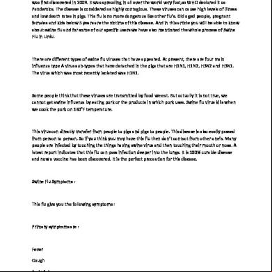
Swine Flu In Urdu 5p4m
December 2021 0
Swine Flu 4s252s
August 2021 0
H1n1 Swine Flu Presentation 23645e
August 2021 0
Swine Production In The Philippines g153l
December 2019 20